Facial Rejuvenation the Entire Spectrum

The quest for youth has spurned a multi-billion dollar industry. Today, the multitude of rejuvenation options is bewildering. A person’s choice of rejuvenation technique is generally determined by his or her physical state, tolerance for pain and downtime, and financial constraints.

The clinical problems that we commonly address include pigmentation, superficial textural imperfections, wrinkles (dynamic or static), deep folds or contour deformities, and skin laxity.

Rejuvenation options are divided broadly into non-invasive (non-surgical) and invasive (surgical) procedures with a few options in the “borderline” zone. Patients should be aware that all these rejuvenation techniques have a common goal of causing controlled damage to the various layers of the skin in order to stimulate repair and regeneration. Sun avoidance and strict usage of sunscreen are the keys to maintaining the results and preventing hyperpigmentation.

NON-INVASIVE (NON-SURGICAL) PROCEDURES

The most common form of phototherapy treatment is Intense Pulsed Light (IPL) or Broadband Light (BBL), which is able to improve the general complexion, lighten pigmentation, improve fine wrinkles and mildly tighten the skin. It is also effective for permanent hair removal. IPL/BBL is a suitable option for regular maintenance without downtime.

To achieve further textural improvement to the superficial layer of the skin, laser resurfacing is more effective. Erbium Yag lasers or carbon dioxide lasers are commonly used to ablate a thin epidermal layer. Erbium Yag is more suited for Asian skin types than carbon dioxide laser. The treated area will be raw and require regular application of antibiotics; the downtime for microlaser peel is around 3 to 5 days, after which a patient can resume putting on makeup.

Various treatment modalities are available to further tighten the dermal layer to for lifting and improvement of static wrinkles. Profractional Erbium Yag (or carbon dioxide) laser, radiofrequency, focused ultrasound and infrared waves can be used to cause thermal injury to the dermal collagen and elastin in a controlled manner. This induces regeneration of new collagen with a skin-firming effect. The clinical results are best assessed 3 to 4 months after the procedure, though most patients would notice improvements after about a month.

Botulinum toxin injection is the standard treatment for dynamic wrinkles – i.e., wrinkles that are worsened by facial animation. Effects usually last for 3 to 4 months. Mild bruising may be present at the needle entry sites. Clinical judgement needs to be exercised to deliver the optimal amount of toxin to alleviate wrinkles without causing an unnatural mask-like appearance.
Filler is used to replace volume; it can correct static deep facial folds (e.g., tear troughs and nasolabial folds) and contour deficits (e.g., sunken temporal areas), and can volumise the cheeks for a more youthful appearance. Filler provides an alternative for those patient who wants augmentation of the nasal dorsum without undergoing rhinoplasty. However, very rarely, the injection of filler into the nose can result in blindness or tissue necrosis. The chemical composition of fillers can be biological (e.g., hyaluronic acid or collagen) or synthetic (e.g., polytetrafluoroethylene). Synthetic fillers may last for more than a year but there is risk of infection and granuloma formation. In the interest of safety, shorter-acting filler material is preferable.
Botox and fillers complement each other, and some patients require both for optimal outcome.

INVASIVE (SURGICAL) PROCEDURES

Fat transfer (grafting) has gained much popularity over recent years as a method of volume replacement. Fat is a good filler option because it is autologous and contains a small amount of stem cells with rejuvenating properties. Fat grafting has been shown to improve the healing of post-irradiated chronic wounds. The final result is firmer skin with increased volume and improvement in wrinkles; the procedure may be repeated for a better cumulative result.

A thread lift procedure provides added levels of skin lifting to the cheeks and temporal area. Several threads made of absorbable material are inserted skin deep and stabilised by suturing to the temporal area. Swelling and bruising can last for a few weeks. The initial appearance may appear over-corrected; this normally improves over a few weeks to months. The clinical effects may last for one to two years.

Common surgeries for facial rejuvenation include blepharoplasty (eyelid surgery), brow lift, temporal lift, facelift and neck lift. These procedures can be done as a single procedure or as a combination, depending on the clinical state.

Upper eyelid surgery (blepharoplasty) can be done to create or improve the configuration of double eyelid or remove excessive droopy skin. It is therefore suitable for young patients who wish to create upper lid folds or older patients with droopy and puffy eyelids.

Ptosis (drooping) of the upper eyelid can co-exist with excessive skin. Not all patients with excess skin have ptosis. The various methods of ptosis correction depending on severity include levator plication or advancement, and in more severe cases something referred to as the FOOM flap.

Eyebags are caused by excessive fat accumulation and also laxity of skin and the orbital septum containing the bag of fat. Lower blepharoplasty can address both eyebags and excessive lower lid skin. There are two methods to remove eyebags: subciliary lower blepharoplasty (external method) and transconjunctival lower blepharoplasty (scarless method).

Subciliary lower blepharoplasty is suitable for patients with excess lower eyelid skin and fat pocket. Removal of fat would result in the gathering of excessive loose skin. Transconjunctival lower blepharoplasty is suitable for young patients with no skin laxity/ excess. The fat pocket is removed via a small cut in the back of the lower lid leaving no scar on the exterior.

A facelift and neck lift would address the problem of a sagging chin and jowl. The contour of the neck can be further improved by dividing the muscle band of the neck (platysmal band division for turkey neck deformity).

Endotine facelift is done via small incisions in the hairline, into which Endotine fixation devices are inserted. These are absorbable strips with small claws to lift up the cheek, jowl and neck. They are absorbed in about 3 to 6 months. Thus, Endotine lifting is suited for patients who do not have significant skin excess.

I often counsel patients that surgery to rejuvenate one area of the face may cause the neighbouring non-operated structure to appear out of sync. For example, in severe ageing eyelids, surgery to improve the eyebags without an upper eyelid procedures may cause the upper lid to appear slightly out of place. It is essential to understand that all features of the face age together. To achieve a harmonious appearance, a combination of procedures may need to be done simultaneously to address the various ageing features.

Facial Rejuvenation – From the simplest to the most elaborate, there is an option for everyone

What happens during the ageing process?

The skin becomes thinner with loss of ground substance, collagen, elastin fibres and, occasionally, fat. This is accompanied by uneven accumulation of pigments. The result is deterioration of skin texture and colour, wrinkles, skin laxity and loss of volume.

How do you formulate a rejuvenation regime?

Every patient requires a personalised regime.
I identify the most significant problems and the ones that bother the patient most. I plan the regime based on the patient’s work schedule, tolerance for downtime, pain threshold, risk adversity and financial constraints.

What are the simpler rejuvenation techniques?

For improvement of skin texture, pore size, pigmentation and fine wrinkles, I recommend intense pulsed light (IPL) or broadband light (BBL). This treatment is well tolerated and has no down time. Clinical results are usually seen within two to three treatments up to one month apart. Some patients report results as early as one week after treatment.
Botulinum toxin injection is the standard treatment for wrinkles which are worsened by facial animation. It can improve crow’s feet, frown lines and forehead wrinkles and its effects usually last for three to four months. Clinical judgement is needed to alleviate wrinkles without causing an unnatural mask-like appearance.
Filler replaces volume to correct deep facial folds and volumise the cheeks for a more youthful appearance. For non-surgical augmentation of the nose, fillers can be a temporary alternative. Most fillers are composed of hyaluronic acid which lasts for about six months. Long-lasting filler materials are associated with side-effects such as infection and granuloma formation.
Botox and fillers complement each other and some patients require both.
Several non-invasive therapies are available to tighten the skin moderately. These include the use of infrared wave therapy (Sciton SkinTyte®), radiofrequency (Thermage®), and focused ultrasound (Ultherapy®). Post-procedure oedema and erythema commonly resolve within a few days, but clinical results may not be as dramatic as ablative laser procedure.
Microlaser peel (resurfacing) can further improve superficial skin texture, and the patient can put on make-up about three to five days after the procedure.

What of moderately complex techniques before surgery?

Profractional laser is used to achieve further skin tightening. This ablates narrow channels into the skin, and is used to treat atrophic acne scars. The healing process is faster and patients can return to work in a few days.
Thread lift provides added skin lifting to the cheeks and temporal area. Several barbed threads made of absorbable material are inserted skin-deep and stabilised by suturing to the temporal area. The initial appearance may appear over-corrected but normally improves over a few weeks to months. The effects can last for one to two years. Fat injection (grafting) has become popular for volume replacement. Fat tissue contains stem cells with rejuvenating properties which are harvested from the abdomen or thighs and processed before being injected. Post-procedure swelling and bruising last for a few weeks, and the final result is firmer skin with increased volume and improvement in wrinkles.

How do you devise a surgical plan for facial rejuvenation?

Ageing affects each part of the face at different speeds and times and this must be understood in the context of the desire for facial rejuvenation.

Labiaplasty: Labia Reduction Surgery In Singapore

WHAT IS LABIAPLASTY?

  • Labial skin excess can result from childbirth or the ageing process. The labia minora becomes floppy and protrudes significantly beyond the labia majora. This can result in discomfort and rash. It can be a source of embarrassment while wearing tight-fitting clothing.
  • Labiaplasty is a surgical technique to fashion and remove excess labial tissue. This labia reduction surgery is usually done for aesthetic or functional purposes.
  • A grading system for labia minora hypertrophy has been proposed by David and West (2008):
    • None: The labia minora are concealed within or extend to the free edge of labia majora.
    • Mild/Moderate: The labia minora extend 1-3 cm beyond the free edge of the labia majora.
    • Severe: The labia minora extend >3 cm beyond the free edge of the labia majora.Labiaplasty (Labia rejuvenation or vaginal lip reduction) is a surgical technique to fashion and remove excessive labial tissue.
  • According to a 2014 survey report by the American Society of Aesthetic Plastic Surgeons, there was a 49% increase in labiaplasty procedures done from 2013, making this one of the fastest growing cosmetic procedures. Thus, it is no wonder why an increasing number of people are seeking for the best labia reduction surgeon in Singapore.
Techniques of Labiaplasty
Trim techniqueWedge techniqueZ-Plasty techniqueDeepithelialization technique
Diagram
Labiaplasty - Trim method Labiaplasty – Trim
Labiaplasty - Wedge Excision Method Labiaplasty – Wedge Excision
Labiaplasty - Z Plasty Method Labiaplasty – Z Plasty
Labiaplasty - De-epithelialization method Labiaplasty – De-epithelialization
AdvantagesSimplest, often preferred technique.Maintain the natural mucosa and skin edge. Minimal scarring.Variation of wedge technique. Maintain the natural mucosa and skin edge. Minimal scarring.Nerve and lymphatic preservation
DisadvantagesNerve end interruptionPotential damage to nerves along the edge of the removed wedge. Suitable for only minimal labial excess.Suitable for only minimal labial excess.Limited amount of tissue removed and risk of Recurrent labia hypertrophy


Associated Procedure
Clitoral UnhoodingLabia Majora ReductionG-spot AugmentationMonsplasty
Reduction of the tissue around clitoris to improved appearance and sensitivityResection of excessive labia majora tissue to provide a more harmonious resultInjection of fat grafting and fillers to the G Spot to enhance sensationWedge excision of mons to reduce volume. Can be done with labiaplasty or abdominoplasty.

SUITABLE CANDIDATES FOR LABIAPLASTY

  • A patient affected by rash and discomfort due to excessive labial tissue.
  • A patient who requests enhancement of genital appearance

PROCEDURES DONE TOGETHER WITH LABIAPLASTY


DESCRIPTION OF LABIAPLASTY PROCEDURE

  • Pre-operative markings to determine the amount of excessive labial minora tissue.
  • Administration of sedation and local anaesthesia.
  • Excision along free edge of labial minora to remove excessive labial tissue.
  • Wound closure with absorbable sutures (no removal of sutures needed).
  • Surgical duration : 1-2 hours.
  • Home on the same day.


ANAESTHESIA

  • General anaesthesia
  • Combination of local and intravenous sedation


POST OPERATIVE CARE

  • Medication: Oral and topical antibiotics, analgesics (pain relief), anti-swelling medication
  • Sanitary padding for 2-3 days for residual discharge.
  • Avoid activities that will exert pressure to the groin for 2-4 weeks.
  • May resume sexual activity after 2 weeks.
  • Follow up 2-4 weeks later.


RISK AND CAUTION OF LABIAPLASTY REDUCTION SURGERY

  • Infection
  • Swelling
  • Bleeding
  • Bruising
  • Asymmetry
  • Reduced sensitivity


If you are looking for labia reduction surgeon in Singapore, look no further than SWENG Plastic Aesthetic and Reconstructive Surgery.

After the consultation, our plastic surgeon will advise on a suitable labiaplasty technique. He would also recommend if laser vaginal rejuvenation (tightening) or G-spot augmentation should be included, since these are top choices for other patients.

Droopy & Sagging Eyelid Surgery – Ptosis Correction in Singapore

WHAT IS PTOSIS?

Eye Cross Section - Droopy Eyelid - Blepharoplasty - Ptosis Correction Eye Cross Section – Double Eyelid – Blepharoplasty – Ptosis
  • Ptosis (blepharoptosis) is the abnormally low (droopy) position of the upper eyelid margin due to a weakness in the lifting mechanism. This mechanism consists of muscle (levator palpebral superioris) and its soft tissue extension (levator aponeurosis)
  • It must be differentiated from dermatochalasis. In this condition, excessively lax upper eyelid skin causes a droopy appearance rather than the true weakness of the lifting mechanism of the eye.
  • Blepharoptosis can also co-exist with dermatochalasis.
  • Patients with blepharoptosis or dermatochalasis has tired looking eyes even though they have adequate rest. They usually use their forehead muscles to assist in lifting up the upper eyelids resulting in persistent frownlines. Thus, seeking droopy eyelid treatment is vital.

Ptosis Vs Dermatochalasis

 BlepharoptosisDermatochalasis
Diagram
Blepharoptosis - Droopy Eyelid - Ptosis Correction Blepharoptosis
Dermatochalasis - Droopy Eyelid - Differentiate from Ptosis Correction Dermatochalasis
MechanismDroopy lid due to weakness in the lifting mechanism of eyelid.Droopy lid due to excessive eyelid skin.
What covers the limbus?Ciliary margin (eyelash margin)Skin edge of redundant upper eyelid skin
Age groupAll ageOlder age
Associated disorderBoth conditions can co-exist
Medical claimsConsidered medical conditionConsidered cosmetic condition

*Limbus – Junction between the cornea and the sclera (the black and the white of the eye)


ASSESSMENT OF BLEPHAROPTOSIS

  • Blepharoptosis is assessed based on two parameters :
    • Degree of blepharoptosis – the severity of blepharoptosis
    • Levator function – the residual lifting ability of the upper eyelid opening mechanism


DEGREE OF BLEPHAROPTOSIS

There are two methods to measure the severity of blepharoptosis

1. Amount of upper lid droop or descent (over the limbus)

Blepharoptosis Assessment - MRD measurement for droopy eyelid Blepharoptosis Assessment – MRD

2. Marginal Reflex Distance

    • Most accurate method to measure degree of blepharoptosis
    • It is a vertical distance between the pupillary light reflex and the eyelid margin.
    • Types of MRD :
      • MRD1
        • Distance from the pupil center to the upper eyelid
        • Blepharoptosis present if MRD1 < 4 mm
      • MRD2
        • Distance from the pupil center to the lower eyelid
        • Lower lid retraction if MRD2 > 6 mm
 Lid Descent over Upper LimbusMRD1
Degree of Blepharoptosis
Ptosis Assessment by Coverage of Limbus - Droopy eyelid Ptosis Assessment by Coverage of Limbus
Blepharoptosis Assessment by MRD for droopy eyelid Blepharoptosis Assessment by MRD
Mild1-2 mm3-4 mm
Moderate3 mm2 mm
Severe> 4 mm1 mm


LEVATOR FUNCTION

  • The levator palpebral superioris is the main muscle responsible for eye opening.
  • To measure the residual lifting ability of the upper eyelid opening mechanism, the patient is asked to look downwards and then upwards while immobilizing the brow. The amount of eyelid movement is called levator excursion and this reflects levator function.
  • The levator function and degree of blepharoptosis determine the type of corrective surgery to be done.


Table of Levator Function Assessment

Levator ExcursionLevator Function
Levator Function Test for Droopy Eyelid - Lid Closed Levator Function Test – Lid Closed
Levator Function Test for Droopy Eyelid - Lid Open Levator Function Test – Lid Open
Good : >10 mm
Fair : 5-10 mm
Poor : 0-5 mm


SUITABLE CANDIDATES FOR PTOSIS CORRECTION SURGERY

  • Patient with established blepharoptosis.
  • Patient with blepharoptosis concomitant dermatochalasis. 


DESCRIPTION OF PTOSIS CORRECTION PROCEDURE

Table of Status of Levator Function and Corresponding Ptosis Correction Procedures

 Levator Function
 Excellent (> 10 mm)Moderate (5-10 mm)Poor (0-5 mm)
Type of Procedure
  • Aponeurotic surgery (Fasanella-Servat procedure, Putterman’s procedure, Mutarde’s Split Level Approach)
  • Mullerectomy
  • Levator advancement or plication
  • External levator resection
  • Frontalis suspension
  • Frontalis Orbicularis Oculi Muscle (FOOM) flap
  • Many surgical procedures have been described for the correction of blepharoptosis tailored according to levator function.
  • Common procedures performed by our surgeon is sagging eyelid correction – levator plication and levator advancement.
  • The levator aponeurosis is shortened by pleating and suturing in levator plication procedure. A portion of the excess aponeurosis is removed and the free edges are overlapped and sutured together in the levator advancement procedure.
  • The incisions and scar for blepharoptosis correction are similar to conventional blepharoplasty. The scar is hidden in the eyelid crease.
  • However, the scope of surgery is vastly different. It is a very delicate surgery that require meticulous dissection of the opening mechanism of the eye and it takes longer duration than conventional blepharoplasty.
  • During the operation patient may be required to sit up and check for symmetry of eye opening several times after correction.
  • Ptosis correction ( droopy eyelid surgery )  can be combined with conventional upper blepharoplasty procedure for :
  • Surgical duration: 2-4 hours
  • Day Surgery (home on the same day)


ANAESTHESIA

  • Combination of local anaesthesia with light intravenous sedation administration:
    • Maximal patient comfort
    • Patient able to participate in checking symmetry of eye-opening during the surgery.
  • General anaesthesia:
    • At the special request of the patient.
    • Result might be affected because of the inability to check for symmetrical eye-opening.


POST OPERATIVE CARE

  • Mild discomfort to be expected includes:
    • Swelling usually resolves in 1 week.
      • In rare instances swelling may take several weeks to resolve.
      • Avoid exertion and rest 30 degree head up to hasten resolution of swelling.
    • Bruising usually resolves in 2 weeks.
    • Tearing
    • Lagophthalmos
      • Incomplete eyelid closure – usually transient
      • May need lubricant eye drop or eye gel to prevent corneal desiccation (drying).
      • Eye pad may be required during sleep.
    • Post-operative medication:
      • Antibiotic ointment to be applied regularly 3-5 times a day
      • Antibiotic eye drop 3 times a day
      • Oral antibiotics
      • Analgesics (pain relief) and anti-swelling medications
    • Removal of suture at 1 week.
    • Resumption of light exercise for 3-4 weeks.
    • Refrain from wearing contact lenses up to 4 weeks post-surgery.


RISK AND COMPLICATION OF DROOPY EYELID CORRECTION TREATMENT

  • Bleeding/ Haematoma (blood clot accumulation)
    • Avoid exertion post-operative.
  • Infection
    • Very rare
    • Avoided by clean (sterile) technique during the surgery.
  • Asymmetrical eye-opening is fairly common in the early post-operative period


FREQUENTLY ASKED QUESTION

Is asymmetrical eye-opening common after blepharoptosis correction?

  • This is a common occurrence and it is usually transient. The eye will take a short while to adjust to its new eyelid opening position.
  • Many patients have pre-existing asymmetrical height of the globe of the eye, the size of eye-opening, the amount of extra skin and fat in the eyelids, and the distance between the brow and upper lid.
  • Due to all these factors, asymmetry can still result despite best efforts during surgery.


Is it common to perform ptosis surgery (sagging eyelid correction surgery) in only one eye?

  • Although blepharoptosis may be unilateral (exist in one eye) it is common to suggest a correction in both eyes because ptosis correction in one eye can affect the eye-opening of the opposite (unoperated) eye. During the consultation, Dr Ng would explain why this is affected by the complex nature of equal innervation of both eyes (Hering’s law).

COST OF DROOPY EYELID SURGERY

  • Blepharoptosis correction is considered medical and financial assistance by medical insurance is possible provided a set of strict criteria are met. These criteria include special test e.g. visual field testing by an eye specialist.
  • The amount of reimbursement by the insurance company is determined on a case-by-case basis by their assessment team.


During your consultation, our plastic surgeon will explain the most common causes of ptosis, sagging or droopy eyelids, and recommend the most suitable droopy eyelid correction surgery and treatment methods for you.

Hymenoplasty (Hymen repair)

INTRODUCTION

  • Hymenoplasty or Hymenorraphy is a surgical reconstruction or repair of the hymen membrane (ring-like skin covering the entrance to the vagina).
  • Intact hymen in some cultures or places is the basis of a woman’s virginity. Blood on wedding sheets after the first sexual intercourse following the marriage is recongised as a proof of a woman’s virginity.
  • The tearing and bleeding of the hymen cannot be used as a genuine confirmation of virginity. Some women tear their hymen membrane during vigorous exercise or due to use of tampons.
  • Various techniques of hymenoplasty:
    • Suturing the torn hymen (true repair of hymen) :
      • This is a very delicate surgery that has to be done with precision and the surgeon must ensure durability of the result.
      • Dr Ng prefers double repair method. A more durable methodology.
    • Vaginal lining flap :
      • Done by cutting a piece of vaginal lining to reinforce the hymen and sometimes this is combined with the injection of a gelatin capsule to fake bleeding during sexual intercourse.


SUITABLE CANDIDATES

  • Patients who require an intact Hymen for personal, cultural or religious purposes.

CONCURRENT PROCEDURES

ANAESTHESIA

  • A combination of local and intravenous sedation (The procedure can be assisted by an anaesthetist according to patient preference).

DESCRIPTION OF PROCEDURE

  • Administration of sedation and local anaesthesia.
  • Identification of torn edges of the hymen followed by a repair using absorbable sutures in two layers.
  • Surgical duration : 1 hour
  • Home on the same day

POST OPERATIVE CARE

  • Medication : Oral and topical antibiotics, analgesics (pain relief), anti-swelling medication
  • Sanitary padding for 2-3 days for residual discharge.
  • Avoid activities that will exert pressure to the groin for 2-4 weeks.
  • Earliest time for sexual activity : 2-3 weeks.

RISK AND CAUTION

  • Infection
  • Swelling
  • Bleeding

Double Eyelid Surgery – Incisional Blepharoplasty

ASIAN VS CAUCASIAN EYELIDS

Caucasian Double Eyelid vs Oriental Single Eyelid - Double Eyelid Surgery Caucasian Double Eyelid vs Oriental Single Eyelid
  • The levator aponeurosis is part of the opening mechanism of the eye and which is closely related to the tarsal plate. It sends multiple slips of dermal (skin) extension which pulls on the pretarsal upper eyelids skin creating the double eyelid fold (palpebral fold).
  • In many Oriental patients, the levator aponeurosis expansions (penetration) into the pre-tarsal crease are absent. This results in single eyelid appearance.

INTRODUCTION

  • Upper eyelid surgery (blepharoplasty) can create or improve the configuration of double eyelid or remove excessive droopy skin. It can be done by incisional method or by suture technique.
  • There are 2 methods of upper eyelid surgery:
  • Purpose of incisional blepharoplasty :
    • Creation of double fold :
      • During incisional blepharoplasty, excess skin is removed and very fine sutures secured the wound edge (which is the location of the double fold) to the deeper eyelid structures to create a permanent fold.
    • Removal of excessive upper eyelid skin (which causes the eye to appear “aged” and may obstruct vision)
    • Removal of excessive upper eyelid fat pad (which gives the eyelid a puffy appearance), eyebag surgery is often done concurrently.
    • Correction of ptosis (Droopy eyelid)

COMPARISON OF SUTURE AND INCISIONAL BLEPHAROPLASTY

Suture Double Eyelid Surgery(scarless)Incisional Double Eyelid Surgery
Photo
Suture Blepharoplasty - Scarless Double Eyelid Surgery Suture Blepharoplasty
Suture blepharoplasty - Cross section - Scarless Double Eyelid Surgery Suture blepharoplasty – Cross section
Incisional Blepharoplasty - Double eyelid surgery Incisional Blepharoplasty
Purpose
  • Creation of double eyelid fold in young patient who have no upper eyelid skin excess or fat excess.
  • Creation of double eyelid fold (for all age groups)
  • Removal of excessive upper eyelid skin
  • Removal of excessive upper eyelid fat pad.
  • Ptosis correction
ScarScarlessFaint scar in the upper eyelid crease
Longevity of ResultLess permanent upper eyelid fold (unpredictable loss of fold)Permanent
Post-surgery downtimeGenerally limited swelling and bruising (however some patients has downtime similar to incisional blepharoplasty)Swelling much improved after 1-2 weeks
Suture RemovalUsually none1 week post operative

STYLE OF UPPER LID DOUBLE FOLD (PALPEBRAL FOLD)

TAPERED FOLD (IN FOLD)PARALLEL FOLD (OUT FOLD)
Upper Eyelid - High Tapered fold - Blepharoplasty - Double eyelid surgery Upper Eyelid – High Tapered fold – Blepharoplasty
Upper eyelid - Parallel fold - Blepharoplasty - Double eyelid surgery Upper eyelid – Parallel fold – Blepharoplasty
  • More common among Orientals.
  • Inner corner of the double fold starts of nearer the eyelash margin and gradually tapers outwards.
  • More common among Caucasians.
  • Entire length of double fold is almost parallel to the eyelash margin.
LOW FOLDHIGH FOLD
Upper eyelid - Low in-fold - Blepharoplasty - Double eyelid surgery Upper eyelid – Low in-fold – Blepharoplasty
Eyelid - Tapered fold (high) - Double eyelid surgery Eyelid – Tapered fold (high)
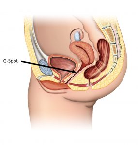
  • Narrow separation of double fold.
  • Appears more natural.
  • Double fold tends to disappear earlier with ageing.
  • Wide separation of double fold.
  • Appears less natural.
  • Results last longer and becomes more natural with ageing
  • The style of double eyelid fold is summarized in the table above.
  • Some patients have strong tendency towards forming parallel fold. It may not be easy to create a tapered fold in this group of patients.
  • During consultation Dr. Ng will advise you regarding the most suitable style and height of double eyelid fold to be created.
  • In some instance the original height of the double eyelid crease can be maintained.

SUITABLE CANDIDATES

  • Patient of any age group who wishes to create double upper eyelid fold.
  • Patient with excessive upper eyelid causing aged appearance and/or obstructing vision
  • Patient with puffy eyelids due to fat pad excess
  • Patient who has weakness in the opening mechanism of the eye (Ptosis)

DESCRIPTION OF PROCEDURE

  • Marking and taping simulation of the intended fold height before surgery in agreement with patient
  • Incision over intended eyelid fold
  • Removal of excessive upper eyelid skin, muscle and fat pads if present
  • Upper eyelid crease is secured by tagging the intended fold to the deeper structures including tarsal plate.
  • Meticulous wound closure with very fine non-absorbable sutures.
  • Topical antibiotic ointment
  • Surgical duration : about 1.5 hours
  • Home on the same day

ANAESTHESIA

  • Combination of local anaesthesia with IV sedation administration – for greatest comfort

POST OPERATIVE CARE

  • Mild discomfort to be expected includes:
    • Swelling usually resolves by 1 week.
      • In rare instances swelling may take several weeks to resolve.
      • Avoid exertion and rest 30 degree head up to hasten resolution of swelling.
    • Bruising usually resolves by 2 weeks.
    • Tearing
  • Post-operative medications:
    • Antibiotic ointment to be applied regularly 3-5 times a day
    • Antibiotic eyedrop 3 times day
    • Oral antibiotics
    • Analgesics (pain relief) and anti-swelling medications
  • Removal of suture at 1 week.
  • Resumption of light exercise for 3-4 weeks.
  • Refrain from wearing contact lens up to 4 weeks post-surgery.

RISK AND COMPLICATION

  • Bleeding/ Haematoma (blood clot accumulation)
    • Avoid exertion post-operative.
  • Infection
    • Very rare
    • Avoided by clean (sterile) technique during the surgery.

FREQUENTLY ASKED QUESTION

Can we ensure symmetrical result after blepharoplasty ?

  • Perfectly symmetrical eyes are uncommon.
  • Many patients have pre-existing asymmetrical height of the globe of eye, size of eye-opening, the amount of extra skin and fat in the eyelids, and the distance between the brow and upper eyelid.
  • Because of all these factors, asymmetry can still result despite best efforts to plan the fold symmetrically.
  • Incisional blepharoplasty is more suitable to improve symmetry if you have obviously asymmetrical upper eyelids.

Can double eyelid surgery give me brighter and more alert looking eyes ?

  • This depends on whether the patient has pre-existing droopy upper eyelids (blepharoptosis)
  • The “bright, alert and non-sleepy” appearance of the eyes is determined by how wide the upper eyelid can open (imagine this as the “window opening”). This opening is controlled by the lifting mechanism of the upper eyelid which is in the deepest layer of the upper lids. In the usual double eyelid surgery, we operate on the superficial layers and not the deeper layer (imagine this as “trimming the curtain” only). Therefore, if the patient has pre-existing droopy upper eyelids (blepharoptosis), simple double eyelid surgery cannot restore the bright and alert appearance.
  • If the patient has no pre-exiting blepharoptosis but only significant skin excess, creation or restoration of double eye fold together with removal of skin excess can potentially brighten up the eyes.
  • This is the reason occasionally double eyelid consultation become prolonged due to additional explanation needed in some cases.

A consultation would guide you regarding which is the most common and best procedure used by top Singapore plastic surgeon for double eyelid surgery.

G-Spot Augmentation

INTRODUCTION

  • G- Spot also known as the Grafenberg spot. It is the erogenous area of the vagina that when stimulated, may lead to strong sexual arousal, powerful orgasms and potential female ejaculation. (Morris, Desmond (2004). The Naked Woman: A Study of the Female Body. New York: Thomas Dunne Books. pp. 211–212. ISBN 0-312-33852-X.)
    G-spot Location G-spot Location
  • It is said to be located within 1-2 inches in the anterior vaginal wall. It feels like a spongy bump which is different from the other part of the vaginal lining.
  • The G-Spot can be augmented to provide better sensation by injection of Hyaluronic Acid filler or autologous fat in this region.


SUITABLE CANDIDATES

  • Females that wants to enhance their sexual gratitude and pleasure.

CONCURRENT PROCEDURES

DESCRIPTION OF PROCEDURE

  • Sedation can be given according to patient’s preference.
  • Fat harvest from inner thigh or abdomen and injected to the G-spot using a very fine needle.
  • Alternatively, a hyaluronic acid filler can be injected.
  • Procedure duration: 15 min – 1 hour.
  • Home on the same day.

ANAESTHESIA

  • Sedation can be given according to patient’s preference.

POST OPERATIVE CARE

  • Medication : Oral and topical antibiotics, analgesics (pain relief), anti-swelling medication
  • Sanitary padding for 2-3 days for residual discharge.
  • Avoid activities that will exert pressure to the groin for 2-4 weeks.
  • May resume sexual activity after 2-3 days.

RISK AND CAUTION

  • Possible minimal bleeding and mild discomfort for 1-2 days

Breast Augmentation: Breast Fillers & Implants Surgery in Singapore

Breast - Anatomy Breast – Anatomy
  • Breast augmentation using implants has been an established procedure with a long historical record of safety.
  • It is the most assured and quickest way to achieve the desired breast volume.
  • The use of implants is one of the best breast enhancement surgery options available in Singapore.

SUITABLE CANDIDATES FOR BREAST ENHANCEMENT SURGERY

Suitable patients for breast implants include individuals with:

  • Mammary hypoplasia or Micromastia (small breast)
  • Significant breast asymmetry
  • Slight breast ptosis
    • After breastfeeding some patients experience significant upper pole breast volume loss. Suitably sized breast implant can provide some lifting effect and restoration of the upper pole volume loss.
  • Post mastectomy reconstruction

PROCEDURES DONE TOGETHER WITH BREAST AUGMENTATION

  • Nipple reduction
    • Enlarged, drooping nipples are commonly encountered especially after breastfeeding. However, many patients often neglect the importance of a matching nipple size when considering breast augmentation.
    • Nipple reduction is essential to achieve a harmonious result.
  • Mommy make-over – abdominoplasty combined with breast augmentation
    • It is common for women after childbirth to experience both deflation in breast volume and lax abdominal skin with stretch marks. Mommy makeover which is a combination of breast enlargement surgery (lift and implant) with abdominoplasty (tummy tuck) is a common request. This combination treatment provides significant rejuvenation effects to the entire torso.
  • Mastopexy
    • This is a surgery for patients with significant drooping of breast tissue. Breast implants can be inserted to provide additional volume and lifting effects.

WHAT DO I NEED TO CONSIDER WHEN UNDERGOING BREAST AUGMENTATION SURGERY?

  • Here are some of the most important factors to be considered:
    1. Size of the Breast Implant
    2. Type of Breast Implant: Saline vs Silicone
    3. Shape of the Breast Implant
    4. Surface of the Breast Implant
    5. Surgical Incision
    6. Placement of the Breast Implant
  • Based on Dr. Ng’s experience with Asian patients, the most common request is for teardrop shaped silicone breast implants, sized between 200-300ml placed via inframammary incision into the sub-pectoral pocket.

1. SIZE OF THE BREAST IMPLANT

  • The size of the breast implant should be in proportional to body stature, height and chest circumference.
  • It is also determined by the width of the breast base, the amount of breast tissue and skin elasticity.
  • Placing excessively large implants will result in an unnatural appearance if there is insufficient tissue coverage.
  • During the consultation, you can try a variety of implant sizes to simulate the final outcome.

2. TYPE OF BREAST IMPLANT: SALINE VS SILICONE IMPLANTS

Silicone Breast Implant - Cohesive Gel Silicone Breast Implant – Cohesive Gel
SalineSilicone
Popularity
  • Less popular
  • More popular
Texture
  • Less natural feel
  • More natural feel
Shell Wrinkling
  • Common
  • Rare
Implant Volume
  • Adjustable
  • Fixed
Adjustment to body temperature
  • Adjust quickly
  • Adjust slowly (e.g. the implant remains cold for a longer duration after a swim)
Contracture Rate
  • Low
  • Higher
Leakage
  • Leakage causes complete deflation but safely absorbable
  • Leakage may cause local inflammation and granulomas, but much safer for modern cohesive gel implants

3. SHAPE OF THE BREAST IMPLANT

Round/ CircularAnatomical/ Teardrop
Profile
  • Dome shaped – less natural appearance
  • Tear drop shape – more natural appearance
  • Large variety of shapes based on different height and projections
Implant Orientation
  • Less crucial
  • Very crucial
Incision
  • More options for incision
  • Usually inframammary incision

4. BREAST IMPLANT – SURGICAL INCISIONS

Breast implant : Choice of Incisions Breast implant : Choice of Incisions
InframammaryPeriareolarAxillaryTrans-umbilical
Scar
  • Scar well-hidden within breast fold
  • Scar at areolar margin – may result in hypertrophic scars
  • Hidden in the armpit crease but visible when wearing sleeveless
  • Well hidden in the umbilicus
Accuracy of implant placement
  • Direct visualization of orientation of implant
  • Best accuracy for all implant placement
  • Reasonably accuracy of implant placement
  • More difficult to ascertain the orientation of implant
  • Least accurate placement of implant
Limitations on implants
  • No limitations
  • Limit choice of implant size if areolar diameter is < 3.5- 4 cm
  • Difficulty with teardrop shape implant
  • Suitable only for saline implant not for silicone implants
Breastfeeding
  • Not affected
  • Can be affected
  • Not affected
  • Not affected
Remarks
  • Most popular approach for tear drop shape silicone implant
  • May cut through breast tissue to allow implant placement
  • Risk of nipple numbness
  • Risk of skin numbness around armpits and upper arm
  • Difficult to lower inframammary fold if needed
  • Difficult and blind dissection
  • High or asymmetrical implant placement

5. SURFACE OF THE BREAST IMPLANT

TexturedSmooth
Contracture Rates
  • Lower
  • Higher
Implant Migration
  • Infrequent
  • Common
Shell Thickness
  • Thicker – maybe palpable
  • Thinner – less palpable

Polyurethane (PU) -covered implant – had a very low contracture rate, but has been withdrawn because of a carcinogenic compound from PU breaking down (although at very low levels).

6. PLACEMENT OF THE BREAST IMPLANT

Sub-glandularSub-pectoral
Diagram
Breast implant - Sub-mammary placement Breast implant – Sub-mammary placement
Breast implant - Sub-pectoral placement Breast implant – Sub-pectoral placement
PlacementImplant underneath breast tissue, above pectoris major muscle – is more anatomicalImplant underneath pectoralis muscles – is less anatomical
Suitable CandidatesFor patient with sufficient original breast tissue – result can be very naturalFor patient with very little breast tissue –pectoralis major muscle provides additional coverage to camouflage edge of implant.
Natural AppearanceMore natural appearance  if there is sufficient breast parenchymalBetter appearance for those patients with very little breast parenchymal
Capsular ContractureHigher riskLower risk
Nipple SensationMaybe affectedNot affected
Implant Size LimitationAllow larger implantImplant size limited by size of subpectoral pocket
Limitation of activityNot likelyChest muscles contraction may cause implant to shift laterally (“dancing breast”)
RecoverySlightly less painSlightly more pain
  • It is advisable to consult an experienced breast enlargement surgeon before proceeding with the surgery.

DESCRIPTION OF BREAST ENLARGEMENT SURGERY PROCEDURE

  • Pre-operative sizing during the consultation :
    • Patient to try multiple implant sizes to simulate the final outcome.
  • Pre-operative screening includes a blood test and breast imaging (ultrasound, mammogram)
  • In a typical surgery via inframammary incision :
    • Pre-operative marking includes planning of the new inframammary fold and incision.
    • Incision at the new inframammary fold
    • Dissection to create space either a sub-glandular or sub-pectoral pocket for implant placement.
    • Insertion of the implant.
    • Meticulous wound closure in multiple layers.
  • Support bra are worn immediately post-operatively.

KEY POINTS IN BREAST FILLERS SURGERY

  • Accurate assessment of suitable implant size. The size of the implant has to be proportional to a patient’s body stature, height and chest circumference. It is also determined by the width of the breast base, the amount of breast tissue and skin elasticity.
  • Accurate placement of the new inframammary fold.
    • Most patients undergoing breast fillers surgery have insufficient breast tissue and tight skin envelope. The inframammary fold needs to be lowered to accommodate the implant to avoid a high riding appearance.
    • It is crucial to determine how much the inframammary fold is to be recited according to the implant size and elasticity of the native skin envelope.
  • Accurate dissection of implant pocket :
    • The size of the implant pocket determines the final appearance of the implant. An inadequately dissected pocket can cause rippling of the implant surface. Over dissection can result in implant displacement and synmastia (bilateral breast pockets communicating together with loss of cleavage).

ANAESTHESIA

  • Dr Ng prefers general anaesthesia for greater patient comfort and safety.
  • In order to create a sub-pectoral pocket for insertion of the implant, the chest muscles need to be relaxed. This may compromise the patient’s breathing capability if done under deep sedation. General anaesthesia is safer because continuous oxygen delivery to the patient is ensured throughout the surgery.

WHAT TO EXPECT POST OPERATIVELY

  • Home on the same day
  • Medication includes antibiotics, pain relief and medication to reduce swelling and bruising.
  • Support bra to prevent the implant from displacing laterally or riding high. The patient wears this bra for at least a month depending on the clinical outcome.
  • Back to work (non-strenuous) within 2-3 days.
  • Stitches (if any) are removed in about 2 weeks.
  • Breast massage:
    • Initiated from 2-3 weeks onwards to further improve the final appearance.
    • Usually the direction of massage is downwards and inwards. This may differ depending on clinical status.
  • Scar management:
    • Scar massage to soften the scar
    • Scar gel application to prevent scar thickening and reduce redness.
    • Photo Therapy to further reduce pigmentation.
  • Exercise:
    • Light exercise can be restarted after 1 month.
    • Avoid exercise that involved heavy usage of the pectoralis muscles for at least 2 -3 months.
  • Appearance of breast:
    • It is normal to expect swelling and tightness over the breast for the first 1-2 weeks. During this period of time the implant may feel more stiff and firm. The breast tissue covering the implants stretches over the next 2-3 months, the implants will feel softer and more natural.
    • Final appearance is usually achieved from 3 months after the breast enlargement surgery. 

RISK AND COMPLICATION OF BREAST SURGERY

  • Bleeding and infection are uncommon.
  • Infection is kept to a minimum by the use of antibiotics, good surgical techniques and having the surgery done in a sterile operation room.
  • Synmastia
    • Is a condition where both breast tissues were merged across the sternum due to over dissection during breast augmentation.
  • Low risk of implant rupture due to modern day implants.
  • Implant displacement
  • Seroma
  • Capsular contracture

COST OF BREAST AUGMENTATION

  • Breast reconstruction using implant post mastectomy due to breast cancer may be considered as a non-cosmetic procedure.

FREQUENTLY ASKED QUESTIONS

Are silicone breast implants safe?

  • Silicone breast implants have been used for several decades for cosmetic as well as reconstructive purpose. They have a good safety record despite being temporarily banned for cosmetic usage in the United States for possible association with connective tissue disease. This was found to be untrue and the ban was lifted.

What is the biggest implant for me?

  • The size of the implant has to be proportional to the patient’s body stature, height and chest circumference. It is also determined by the width of the breast base, the amount of breast tissue and skin elasticity. The placing of excessively large implants will result in an unnatural appearance if there is insufficient tissue coverage.

What is the length of the scar?

  • In most cases, the scar ranges from 3.5 to 4.5 cm depending on the size of the implant chosen. It would be unwise to place a very large implant through the narrowest incision – implant rupture during surgery rarely occurs. During the consultation Dr Ng shall illustrate this point.
  • Through a very small incision, the dissection of the implant pocket and the control of bleeding (haemostasis) is much more difficult.

Can I achieve a natural cleavage after breast augmentation?

  • During dissection of the implant pocket, it is conventional to keep about a 3 cm wide spacing of central chest tissue intact. This is to prevent over dissection which can result in the breast pockets from both sides communicating with each other (synmastia). This is a very difficult complication to rectify.

Do I need to replace the implants after many years?

  • The silicone implant theoretically can last indefinitely i.e. the silicone in the implant does not harden over time. As long as there is no significant capsular contracture there is no need to replace the implant.

What is capsular contracture?

  • The body naturally produces a thin membrane (capsule) around any implanted foreign object, this membrane normally remains thin and pliable. In some patients, significant scarring occurs in the membrane in this capsule, causing the whole construct to appear stiff and hardened (capsular contracture).
  • The severity of capsular contracture has been classified by Baker (1980)
    BAKER CLASSIFICATION SYSTEM OF CAPSULAR CONTRACTURE
    Grade INo palpable capsuleThe augmented breast feels as soft as an unoperated one.
    Grade IIMinimal firmnessThe breast is less soft and the implant can be palpated, but is not visible.
    Grade IIIModerate firmnessThe breast is harder, the implant can be palpated easily, and or implant outline (or distortion from it) is visible.
    Grade IVSevere contractureThe breast is hard, tender, painful, and cold. Distortion is often marked.
    Usually implant replacement is done for grade III to IV capsular contracture.

Can I breastfeed after breast augmentation?

  • Breast enhancement surgery should not interfere with breastfeeding as long as milk ducts are not cut. Other than peri-areoalar incision there is a very low chance of affecting breastfeeding.

Can I still do breast screening examinations?

  • After breast augmentation, cancer screening can still be performed using ultrasound, mammography, and MRI scan. There are special mammographic views (Eklund Displacement) available for better visualization after breast augmentation.

If you are looking to undergo breast enhancement surgery in Singapore, SWENG Plastic Aesthetic and Reconstructive Surgery is the place for you. During your consultation, Dr Ng will explain the common breast enhancement options. He will recommend the most suitable breast enhancement surgery methods used by some of the top breast enlargement surgeons, including those in Korea.

Abdominoplasty (Tummy Tuck Surgery)

INTRODUCTION

  • Weight gain and post-child often result in excessive abdominal skin, fats and skin striae. Liposuction is useful in removing excessive fats in young patients with good skin elasticity. However for older patients with poor skin elasticity, liposuction may result in excessive unsightly loose skin.
  • Abdominoplasty (Tummy Tuck) is a definitive way to remove excessive skin and fat from the abdomen. It is particularly helpful in removing post childbirth abdominal skin excess with stretch marks (striae).
  • The rectus abdominis muscle (the 6 pack muscle) can be overstretched during pregnancy leaving a gap between them (divarication of recti). This gives the abdomen a very full appearance even if there is little excess fat.
  • During this procedure, any abdominal muscle separation (divarication of recti) will also be tightened (plication of divarication of recti), resulting in a flatter abdomen and more of an hour glass waistline.
  • To further improve the result liposuction of the abdomen can be done during the same surgery.
  • Occasionally abdominoplasty can also be combined with abdominal hernia repair. Abdominal hernia is a result of bulging of abdominal contents through the weakness of the abdominal wall.
  • Abdominoplasty is a procedure that can radically contour the abdomen at the expense of having a reasonably well hidden scar. There are many patients who cannot accept having an abdominal scar and who are willing to live with a less than ideal abdominal contour.

TYPES OF ABDOMINOPLASTY:

Conventional AbdominoplastyMini AbdominoplastyFleur De LisBelt Abdominoplasty
Diagram
Abdominoplasty - Conventional Tummy TuckAbdominoplasty – Conventional Tummy Tuck
Abdominoplasty - Mini Tummy TuckAbdominoplasty – Mini Tummy Tuck
Abdominoplasty - Fleur De Lis Tummy Tuck Abdominoplasty – Fleur de Lis
Abdominoplasty - Belt Tummy Tuck Abdominoplasty – Belt
PurposeRemoval of massive abdominal skin and fat; and plication of divarication of recti.Removal of minimal to moderate abdominal skin and fat just below the umbilicus.Removal of the massive upper and lateral abdominal skin and fat. ie. removal of vertical and horizontal abdominal excessRemoval of massive abdominal, hip and back excessive skin and fat.
IncisionAbove pubic hairline, slightly extended to the hip bones.Short incision above pubic hairline.Above pubic hairline, slightly extended to the hip bones and vertical extension upwards.Circumferential incision from front to back.
Umbilical RepositioningUmbilicus repositioned as the abdominal skin pulled downwards. Small scar around the umbilicus.Umbilicus left intact as abdominal skin pulled down. It may be separated from underlying attachment.Similar to Conventional AbdominoplastySimilar to Conventional Abdominoplasty
Associated ProcedureLiposuction of the love handles and upper abdomen for further enhancement of the abdominal shape.Similar to Conventional AbdominoplastySimilar to Conventional AbdominoplastyLiposuction and buttock lift


CONCURRENT PROCEDURES

Abdominoplasty can be combined with the following :

  • Liposuction and fat grafting to breasts and face
    • Fat can be harvested using a syringe from the abdomen before abdominoplasty. The harvested fat can then be injected into the breast for volume enhancement and into the face for rejuvenation.
  • Mommy make-over – abdominoplasty combined with breast augmentation
    • It is common for women after childbirth to experience both deflation in breast volume and lax abdominal skin with stretch marks. Mommy makeover which is a combination of breast augmentation (lift and implant) with abdominoplasty (tummy tuck) is a common request. This combination treatment provides significant rejuvenation effects to the entire torso.
  • Abdominal Hernia Repair
  • Buttock Lift
    • Can be done with Belt Abdominoplasty
  • Thigh Lift
    • Can be done with Belt Abdominoplasty

SUITABLE CANDIDATES

Patients who have :

  • Excessive abdominal fat and poor skin elasticity.
  • Excessive abdominal skin striae (usually post childbirth).
  • Abdominal muscle laxity (divarification of recti) usually post childbirth.
  • Concomitant abdominal herniation
  • Completed family planning. Female patients who intent to become pregnant in future may decide to defer this procedure.

ANAESTHESIA

  • General anaesthesia

DESCRIPTION OF PROCEDURE

  • Pre-operative marking to determine incision and amount of skin to be removed with agreement of patient.
  • Liposuction of the love handles and upper abdomen for further enhancement of the abdominal shape (if necessary).
  • Excision of the excess skin and fat according to markings.
  • Plication of the divarication of recti (tightening of the abdominal muscles by suturing) if necessary.
  • Insertion of wound drains (plastic tubings) near the pubic hairline for evacuation of residual blood to prevent blood clot accumulation.
  • Closure of the abdominal fat and skin in layers and skin dressing. The patient is at rest in a Jack-Knife position (abdomen is flexed)
  • Surgical duration : 4-5 hours


POST OPERATIVE CARE

  • After the procedure the patient may opt to stay in the hospital ward or hotel room with a private nurse for recuperation.
    • The patient usually goes home by the 3rd or 4th day after surgery.
  • In the 1st and 2nd day after surgery the patient rests in a jackknife position to relieve abdominal wound tension.
  • The patient may be able to ambulate with support from 2nd to 3rd day onwards.
  • Removal of drains usually by second or third day when the drainage decreases to an acceptable level.
  • Abdominal binder : Wear round the clock for about 3 months to provide support and to reshape the abdomen.
  • Medication: Analgesics (pain killers), Antibiotics and medications to reduce swelling and bruising.
  • Suture removal : 1-2 weeks
  • Bruising and swelling : May last for about 2 weeks.
  • Return to exercise : Light exercise permitted at about 4 weeks: heavy exercise at about 3 months.


FINANCIAL COUNSELLING

  • Abdominoplasty for cosmetic purposes is not insurance claimable.
  • There are very few insurance claimable procedures that can be done together with abdominoplasty.


RISK AND CAUTION

  • Infection
  • Bleeding
  • Bruising – usually resolved by 2 weeks
  • Swelling – usually resolved by 2 – 4 weeks
  • Wound dehiscence – may occur in patients who have a tendency of poor wound healing eg. Diabetes, Smokers, Patients on chronic consumption of corticosteroids, and patients with vascular disease.
  • Numbness – over the abdomen within expectation and can last for 6 – 12 months
  • Undesirable scar can be address by injection or simple scar revision.


During the consultation, our plastic surgeon would guide you to choose the best tummy tuck surgery method used most commonly by some of the leading plastic surgeons in Singapore and Korea. Apart from abdominoplasty, Dr Ng would discuss associated procedures like liposuction, fat grafting and mommy makeover.